Medicine
The Science of Lasik
The 15-minute surgery that retires your glasses is really applied optics. Fuller explains focal lengths, diopters and the cornea, and shows how LASIK and PRK sculpt the stroma to fix myopia, hyperopia and astigmatism.
- Published in
- Edition 1, Spring 2022
- Pages
- 6–7
- Licence
- CC BY 4.0
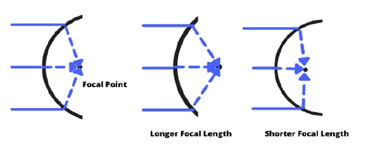
Everyone has heard of LASIK, a 15-minute procedure that promises to eliminate your need for glasses and contacts. Over ten million Americans have gotten LASIK since its FDA approval in 1999, and the numbers have shown it to be widely successful. With such promise, many people are interested in understanding how it works.
At its core, the eye is a two-lens system that works to focus light on the retina. As such, in order to understand how we fix vision, one must understand how optics work in the first place. The guiding principle behind all lenses is refraction, where light bends after entering a new medium. To measure the degree of “bendiness”, or optical power, that a lens imparts on a ray of light, physicists measure the focal length of the lens. A positive focal length converges light, bending it inward, whereas a negative focal length diverges light, bending it outward. A short focal length sharply bends light, whereas a longer focal length does not.
Ophthalmologists use a different unit, the diopter, which is 1/focal length. This unit has a much more intuitive nature; a 3 diopter lens will focus parallel rays of light at ⅓ of a meter away. Therefore, when talking about the eye’s refractive powers, one speaks in terms of diopters.
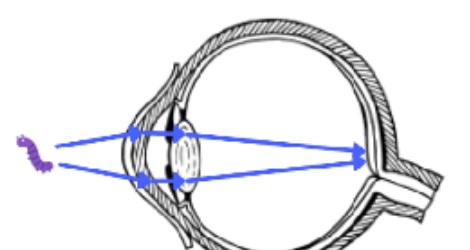
Moving from physics to physiology, let’s imagine a child staring at a bug on the sidewalk. The light reflecting off the bug must first pass through the clear front part of their eye, called the cornea. The cornea is actually composed of three layers: the epithelium (front), the stroma (middle), and the endothelium (back). Seventy percent of the eye’s focusing power, about 43 diopters, actually comes from the cornea, making it an incredibly vital first step in concentrating light. After the light passes through the cornea, it moves through the pupil, and then through the lens. The lens of the eye provides the remaining thirty percent of the focusing power. In a young, healthy eye, the lens undergoes accommodation, in which it changes shape to adjust its refractive power.
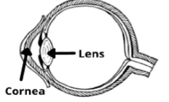
Far-away objects do not require much bending, as their light enters the eye nearly parallel. However, the bug on the sidewalk is quite close, and so the light rays reflecting off of it are much more divergent. To counter this, the child’s ciliary muscles contract, thickening their lens. Once the light is focused, it hits the back of the eye, called the retina, where light-sensitive nerve cells transduce it into electrical impulses. For the purposes of vision correction, the cornea and the lens are the most important structures to note.
While this hypothetical child may have fine vision, a growing issue in the US and around the world are refractive conditions, in which the eye has trouble focusing incoming light. In some people, the eye is either too long or the cornea is too curved. A very steepened cornea would have a low focal length (high optical power), causing light to converge too soon. In either case, light from faraway focuses in front of the retina, causing blurry vision. This condition is called myopia, or nearsightedness. To correct it, a diverging lens is used, which will have a negative diopter measure. For this reason, the severity of myopia is measured in negative diopters. The opposite condition, hyperopia, is categorized by an eye that is too short or a cornea that lacks enough optical power. To correct this, a positive diopter lens is used, and so the severity of hyperopia is measured in positive diopters. A related condition is astigmatism, in which the cornea isn’t perfectly spherical, leading to asymmetry in the optical power. If a regular eye is shaped like a soccer ball, then an astigmatic eye is shaped like an American football. This can cause blurriness or double vision, as images are smeared across different focus points.
These issues are usually corrected through glasses or contacts, but many people opt to permanently change their vision through refractive surgery. Operations like LASIK and PRK, work to modify the shape of the cornea itself. The main difference between them is how they access the strong inner portion of the cornea, called the stroma tissue. The first one to come out was Photorefractive keratectomy, PRK, approved in 1995.
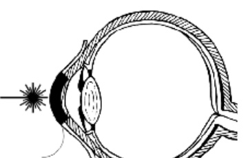
Accessing the stroma is done by removing the epithelium layer using an alcohol solution or a specialized brush. In LASIK, or Laser-Assisted In-Situ Keratomileusis, a flap is cut in the epithelium which is flipped up during surgery, and then flipped back down. Besides the way they access the stroma tissue, the two procedures work in much the same way. They remove sections of the stromal tissue to modify the cornea’s optical power. In myopes, the center of the cornea is flattened, effectively enlarging the focal length, and reducing its optical power. In hyperopes, the periphery of the cornea is removed, shortening the focal length, and enhancing the optical power. Astigmatism involves ablating the asymmetry, rounding out the cornea. This process can be done in conjunction with myopia or hyperopia treatment. These two procedures work remarkably well and can give a patient 20/20 vision within hours after getting off the table.
Ophthalmology is, at its core, applied optics, and so understanding the science behind the eye is essential for both practitioners and patients. Fundamentally, LASIK is simply the reshaping of the cornea, changing or concentrating the focal point in order to improve the image on the retina. Where and how much stroma tissue to remove is based on the pathology of the eye: those who bend the light too much have their focal length increased, and those who don’t bend the light enough have their focal length decreased. The simplicity and effectiveness have rocketed LASIK to the number one refractive procedure in the US.
How to cite this article
Fuller, J. (2022). The Science of Lasik. Columbia Scientist, 1, 6–7. https://columbiascientist.org/articles/science-of-lasik
© 2022 Joshua Fuller. This is an open-access article distributed under the terms of the Creative Commons Attribution 4.0 International licence, which permits use, distribution, and reproduction in any medium, provided the original author and source are credited.