Science Art
My Body Is Art
Watercolor portraits of stem cells, neurons, liver, bone and blood cells float across a black page, a full-bleed collage celebrating the trillions of tiny units that make up a human body.
- Published in
- Edition 2, Spring 2023
- Pages
- 15
- Licence
- CC BY 4.0
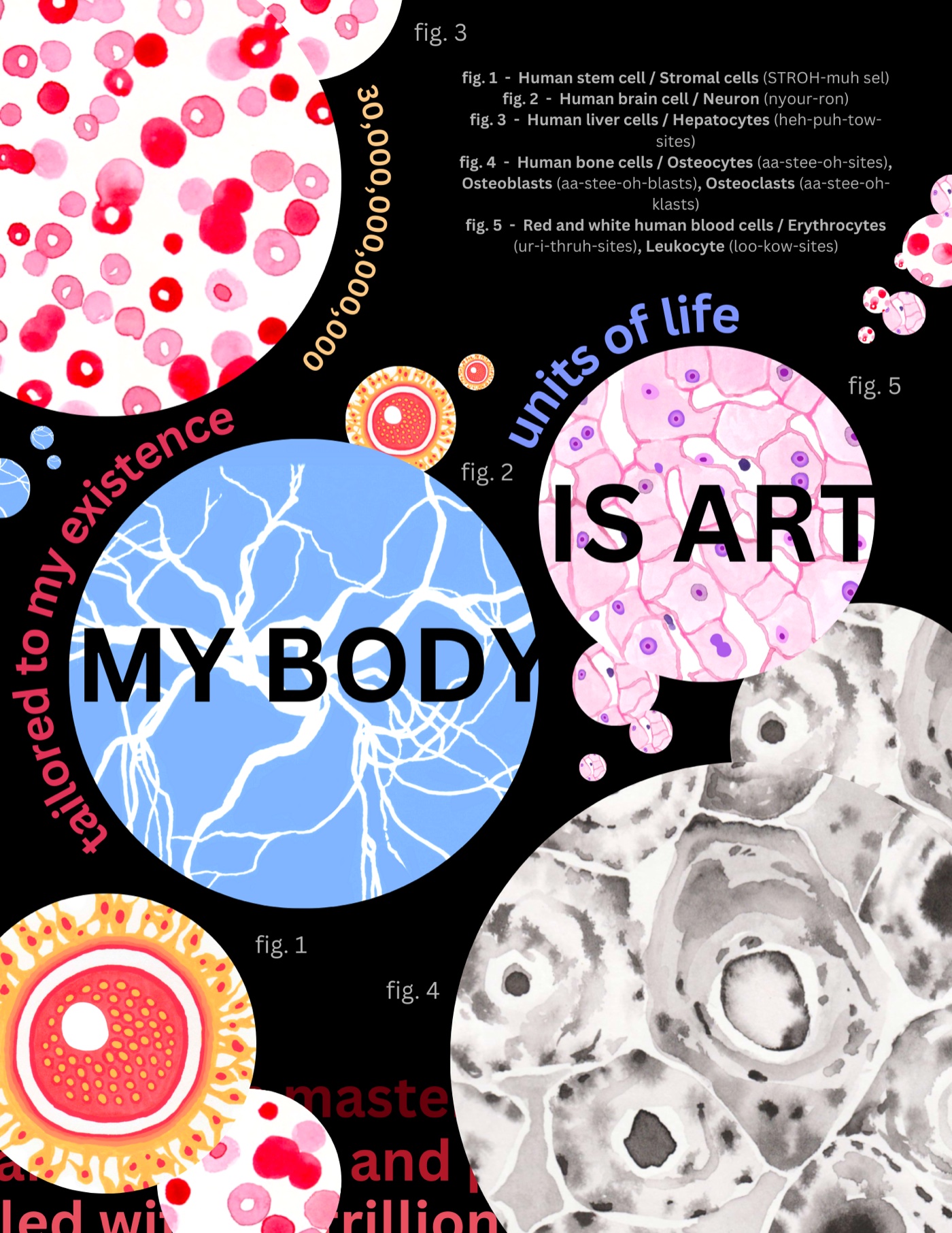
My Body Is Art, a collage of illustrated human cells by Rhiannon Stevens
fig. 1 - Human stem cell / Stromal cells (STROH-muh sel)
fig. 2 - Human brain cell / Neuron (nyour-ron)
fig. 3 - Human liver cells / Hepatocytes (heh-puh-tow-sites)
fig. 4 - Human bone cells / Osteocytes (aa-stee-oh-sites), Osteoblasts (aa-stee-oh-blasts), Osteoclasts (aa-stee-oh-klasts)
fig. 5 - Red and white human blood cells / Erythrocytes (ur-i-thruh-sites), Leukocyte (loo-kow-sites)
How to cite this article
Stevens, R. (2023). My Body Is Art. Columbia Scientist, 2, 15. https://columbiascientist.org/articles/my-body-is-art
© 2023 Rhiannon Stevens. This is an open-access article distributed under the terms of the Creative Commons Attribution 4.0 International licence, which permits use, distribution, and reproduction in any medium, provided the original author and source are credited.